
A practical, clinically grounded guide to recognising, diagnosing, and treating navicular fractures — from cast immobilisation to complex ORIF surgery in Yavatmal.
By Dr. Prashant Kasare, Kasare Hospital
The navicular bone is a small, boat-shaped bone on the top of the midfoot that plays an outsized role in how you walk, run, and bear weight. A fracture here is often missed on first glance because it can masquerade as a simple sprain — yet left untreated, it carries a real risk of long-term pain, arthritis, and deformity. At Kasare Hospital in Shivaji Nagar, Yavatmal, our orthopaedic specialists in Yavatmal regularly manage navicular fractures ranging from simple stress injuries to complex, multi-bone trauma requiring surgical fixation. This article walks through what causes navicular fractures, how to recognise them, and the treatment pathway — including when Open Reduction and Internal Fixation (ORIF) becomes necessary.
The navicular sits between the ankle (talus) and the middle of the foot (cuneiform bones), forming a critical link in the medial arch. It has a unique, limited blood supply concentrated at its centre, with the periphery relying on that central zone — a feature that makes it more vulnerable to poor healing or avascular necrosis (bone death from inadequate blood flow) if a fracture disrupts this supply. This is precisely why navicular fractures are treated with more caution than many other foot fractures: getting the diagnosis and fixation right the first time matters for long-term joint health.
Navicular fractures generally fall into two categories:
Typical symptoms include pain and tenderness over the top of the midfoot, swelling, bruising, and difficulty or inability to bear weight. In stress fractures, pain may build up gradually over days to weeks and worsen with activity rather than appearing suddenly after an injury.
Warning: Seek emergency care immediately if there is visible deformity of the foot, an open wound over the fracture, numbness or a cold, pale foot, or complete inability to bear any weight — these can indicate a complex fracture-dislocation or compromised blood supply that needs urgent assessment.
Diagnosis starts with a clinical examination and plain X-rays of the foot. Because navicular fractures — especially stress fractures and subtle displacement — can be difficult to see on standard X-ray, a CT scan is often required to map the exact fracture pattern, particularly when planning surgery. On-site imaging at Kasare Hospital allows this workup to happen without delay, and intra-operative C-arm guidance helps our surgeons confirm accurate fracture reduction and implant positioning in real time during fixation.
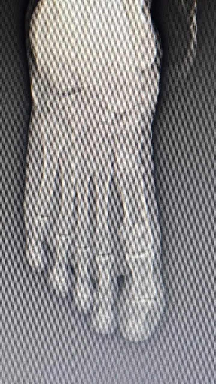
Treatment depends on whether the fracture is stable and non-displaced, or displaced and unstable. Our fracture management team assesses each case individually to decide between conservative and surgical care.
| Approach | When Used | What It Involves |
|---|---|---|
| Conservative (cast/boot) | Non-displaced, stable fractures | Non-weight-bearing cast or boot for 6-8 weeks with regular X-ray monitoring |
| ORIF Surgery | Displaced fractures or joint involvement | Open reduction and fixation with screws or plates to restore anatomy and joint alignment |
| External Fixation | Severe fracture-dislocations with soft tissue compromise | Temporary distraction frame to realign bones before or alongside internal fixation |
For displaced navicular fractures, ORIF using headless compression screws is often preferred because the screw head sits buried below the bone surface, avoiding irritation of the overlying joint cartilage while still delivering strong, stable compression across the fracture. Where surgeons at our hospital also manage related joint injuries requiring arthroscopic surgery, the same minimally invasive philosophy — precise correction with minimal soft-tissue disruption — guides our approach to complex foot trauma as well.
Not every navicular fracture is a straightforward break. One of the more demanding presentations our doctors encounter is a complex navicular fracture with talonavicular joint dislocation and an associated calcaneal fracture — essentially, three connected problems in one foot. In such a case, the fracture cannot simply be screwed back together; the dislocated joint first needs to be gradually realigned before definitive fixation. This was managed using distraction osteosynthesis with a JESS (Joshi's External Stabilisation System) external fixator to carefully restore joint alignment, followed by fracture fixation with headless compression screws once the bones and joint were correctly positioned. This staged approach — external fixation for alignment, then internal fixation for stability — reflects the level of surgical planning that complex, multi-bone foot trauma demands, and is carried out in our International Standard Modular Operation Theatre, the first of its kind in Yavatmal and third in Central India.
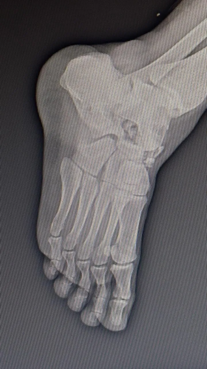
Tip: If your foot fracture involves more than one bone or a visible joint deformity, ask your surgeon specifically about the fixation strategy — staged treatment (external fixation first, internal fixation later) is often safer than a single-stage surgery for complex, multi-bone patterns.
Patients often ask about the \"success rate\" of foot fracture surgery, but outcomes are shaped by several specific factors rather than a single number: how displaced the fracture was, whether the joint surface was involved, how quickly surgery was performed after injury, how well the navicular's limited blood supply was preserved during fixation, and how consistently the patient follows the post-operative weight-bearing and rehabilitation protocol. Delayed treatment or inadequate fixation raises the risk of non-union, malunion, or secondary osteoarthritis of the midfoot joints. Dr. Prashant Kasare brings 15+ years of orthopaedic practice, including foot & ankle surgery training and trauma fellowship experience gained across institutions such as AIIMS, RML Hospital, and Lilavati Hospital, to the planning of each fixation strategy — matching the surgical technique to the specific fracture pattern rather than applying a one-size-fits-all approach.
Recovery from navicular ORIF typically involves a period of strict non-weight-bearing in a cast or boot — commonly 6 to 8 weeks — followed by gradual, protected weight-bearing as X-rays confirm healing. Stiffness and muscle deconditioning are expected after this period of immobilisation, which is why structured physiotherapy is a core part of recovery rather than an afterthought. At Kasare Hospital, integrated physiotherapy is built into the treatment pathway so that rehabilitation begins as soon as it is safe, supporting a smoother return to walking and normal activity.
Kasare Hospital, located in Shivaji Nagar near Shivaji Park, Yavatmal, is built specifically around orthopaedic and trauma care under NABH-standard protocols. Our infrastructure includes an International Standard Modular Operation Theatre, on-site X-ray and diagnostic imaging, an in-house pharmacy, and 24/7 emergency services (with Sunday availability from 9 AM to 2 PM) — meaning a patient with a midfoot fracture can be assessed, imaged, and, if needed, taken to surgery without being referred elsewhere. Since 2014, the hospital has treated over 75,000 patients and performed more than 7,865 admissions and surgeries, reflecting our broader commitment to our quality care standards. You can review our full range of specialities and our diagnostic and surgical technology before your visit.
Any midfoot pain that persists beyond a few days, worsens with weight-bearing, or follows a fall, accident, or sudden increase in running or sports activity should be evaluated promptly rather than \"waited out.\" Early diagnosis is one of the strongest predictors of a good long-term outcome for navicular fractures, since delayed treatment increases the risk of poor healing at this blood-supply-sensitive bone.